Incision-Free Treatment of Urinary Stones
Summary
All female dogs and cats are candidates
Male dogs >10 pounds (can pass a 10 fr urinary catheter)
Stone burden not excessive (too large or too many)
Screen for urinary tract infections, other diseases that would complicate management
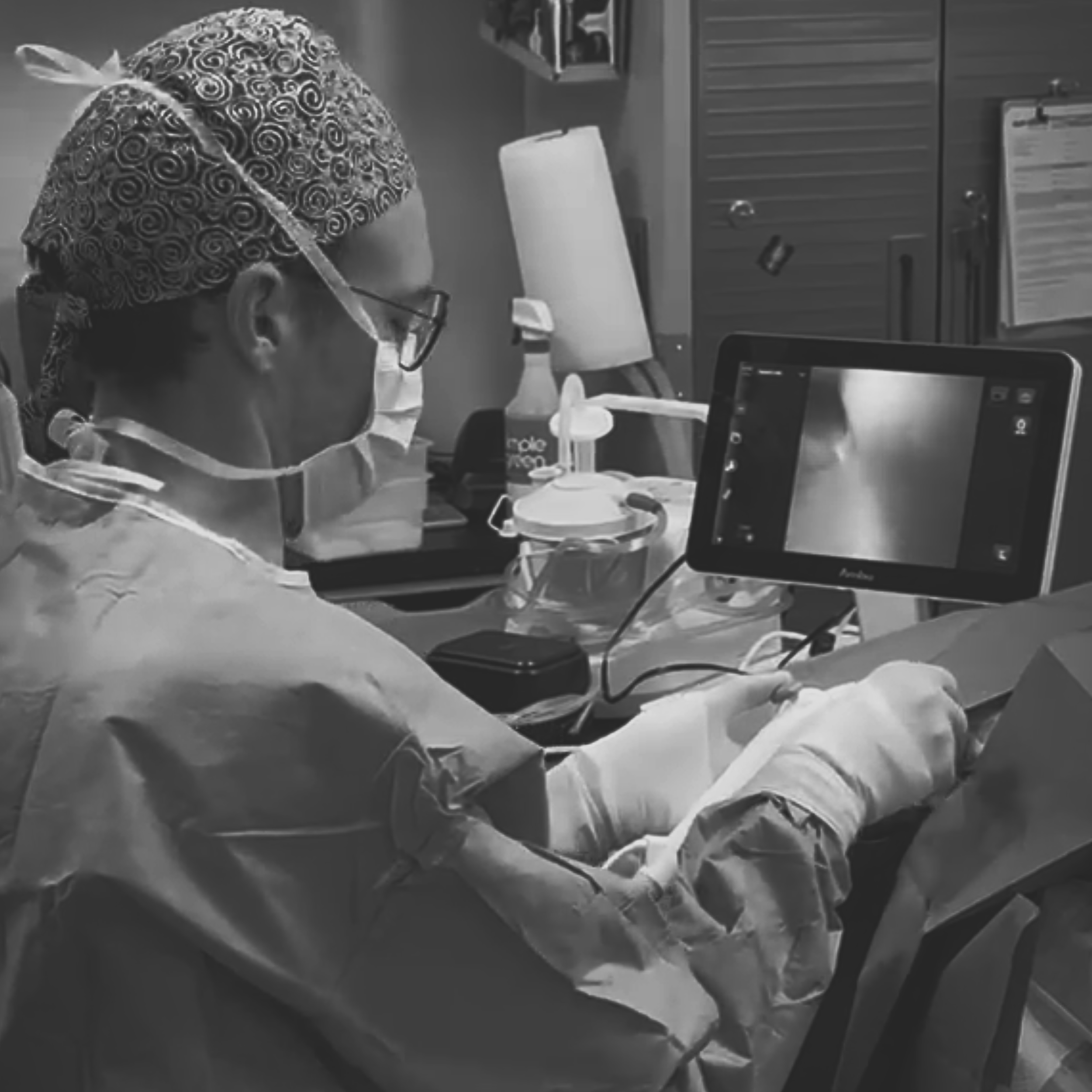
Advanced laser lithotripsy offers a precise, non-surgical approach to treating urinary stones and getting your pet back to a pain-free life sooner.
Redefining Stone Management
Making Medicine Less Invasive
Laser Lithotripsy, when combined with minimally-invasive cystoscopy, offers a less invasive alternative to traditional abdominal surgery. Less invasive procedures result in a faster recovery time because these patients don’t have incisions which means less healing time and less post-operative discomfort. A more traditional open surgical approach is still sometimes needed, but when a patient qualifies for a less invasive option there are numerous benefits.
What is Laser Lithotripsy?
There are many types of lithotripsy technology in medicine which roughly translates from Greek to “stone crushing”. For urinary stones, the use of specialized lasers that can pulverize or fragment stones is most common. These lasers are highly engineered with very specific wavelengths that deliver a burst a very concentrated energy on the stone’s surface creating a vapor bubble, which is called the Moses Effect. When this vapor bubble collapses it causes a jack-hammer like impact on the stone which breaks the stone into smaller pieces.
Multiple laser types have been developed and laser lithotripsy has become the standard of care in human medicine. In veterinary medicine, the technology still isn’t widely accessible, even at specialty hospitals.
The Goal
Over the course of a lithotripsy treatment, the stones are broken into fragments that are small enough to be removed by the scope or be peed out. Ultimately, this means incision-free removal of urinary stones and less discomfort for our pets.
Indications: When to Recommend Lithotripsy
Species and Sex Differences
Female dogs and cats are ideal candidates for cystoscopy and lithotripsy as their anatomy is simpler their urethras are larger. Cystoscopy can also be performed in medium to large male dogs with the right equipment. Male cats, unfortunately, are never a good candidate for lithotripsy unless their anatomy has been previously altered surgically (e.g. perineal urethrostomy).
Ideal Candidates:
Female dogs and cats of all sizes
Male dogs >5kg (must be able to pass 10 fr urinary catheter)
Male cats are not candidates
Urolith Type and Location
Most stones can be treated with lithotripsy but other treatments should also be considered. It’s also important to remember that cystoscopy also has the benefit of offering diagnostic information as well, which may help identify a cause of recurring urinary stones in some pets. Samples of stones and biopsies of the urinary tract can be acquired during cystoscopy, if needed.
Ideal Candidates:
Stones that can’t be medically dissolved (e.g. calcium oxalate)
Stones causing urethral obstruction which would result in a technically difficult surgery
Small to moderate stone burdens in the urinary bladder
Case Selection and Contraindications
Too Big or Too Small?
Cystoscopy can typically be easily performed in almost all female cats and dogs excluding any unusual anatomy or unique circumstances. Male dogs, however, have much narrower and longer urethras and need specialty scopes. Even with specialized equipment, male dogs typically need to weigh at least 10 pounds. Male cats are typically never candidates for cystoscopy.
We also consider something called the “stone burden” which is overall size and number of urinary stones. If there are exceptionally large stones or there are a very large number, the duration of laser lithotripsy procedures can become very prolonged. It’s often faster, and better for the patient, to have traditional open surgery in those cases.
There are still alternatives to open surgery if patients aren’t candidates for cystoscopy, such as the minimally invasive percutaneous cystolithotomy. This is a procedure where a scope is passed into the bladder through a small incision in the abdomen. We’re happy to discuss these options if you have questions!
Medical Contraindications
We always consider whether a patient is a good candidate for general anesthesia as patients need to be anesthetized for lithotripsy procedures. Conditions like underlying heart disease or advanced stages of kidney or liver disease may require additional consideration. Patients with a known history of bleeding disorders or who are prescribed blood thinners may also require additional consultation prior to the procedure.
It’s also ideal to avoid lithotripsy in patients with active urinary tract infections. This isn’t always avoidable, especially when urinary stones are acting as a nidus. If active infection is present, we recommend pre-treating with an antibiotic based on MIC testing for 5-7 days prior to the procedure.
The Procedure: What Happens During Lithotripsy?
Equipment
The equipment used during laser lithotripsy procedures varies depending on several factors including the size of the patient, location of the stones, and overall stone burden. For most patients, a flexible or rigid cystoscope is used which has a small camera and channels that allow us to introduce specialized instruments into the urinary tract.
The type of laser used for lithotripsy is most commonly a holmium:YAG laser, though other types also exist. This type of laser transmits energy down a flexible fiber through the cystoscope which allows direct targeting of urinary stones during the procedure.
A variety of other instruments are sometimes used including small baskets and graspers can be used to remove smaller stones and fragments.
Fragmentation and Dusting
Laser energy effects the various types of urinary stones differently; some stones will tend to crack and break into smaller pieces, which is called fragmentation, while other types of stones erode as the laser energy hits the surface and turns into a fine powder, which is called dusting. Either way, the primary goal of lithotripsy is to break stones into pieces that are small enough to be removed without getting stuck and causing an obstruction.
Final Clearing
Once the pieces are small enough, a combination of voiding and retrieval is typically used to remove all of the remaining stone pieces. Voiding is a processed where the bladder is filled with sterile saline, rinsed, and gently expressed to allow the stone pieces to be flushed out of the urethra. Retrieval uses a variety of baskets and grasping instruments to manually grab and remove the stones using the scope. X-rays are typically used to document that all of the stone fragments have been removed.
The Risks Vs. Benefits
Benefits
No Incision: No sutures, less pain, and immediate discharge (often the same day).
Surgical Precision: Direct visualization ensures all fragments are removed, reducing recurrence from missed "nidus" stones.
Safety: The Ho:YAG laser energy is absorbed within <0.5 mm of fluid, minimizing risk to the bladder wall
Risks
Urethral Edema: Temporary swelling that may require an indwelling catheter for 24–48 hours (rare).
Incomplete Removal: Higher risk in males (20–30%) compared to females due to urethral anatomy.
Iatrogenic Injury: Rare risks of urethral tears, strictures, or bladder perforation (rare).
The Referral Checklist
Abdominal Radiographs: These will evaluate for concurrent ureteroliths or nephroliths which may require additional testing such as ultrasound. We will also evaluate overall stone burden. Please send us radiographs directly; we’re happy to consult on potential cases!
Urinalysis ± Urine Culture: A urinalysis may give clues about the type of stone present, but, more importantly, will assess for overt pyuria. If present, a urine culture should be performed to determine the most appropriate antibiotic prior to the procedure.
Other Lab Testing: Routine lab testing with complete blood count and serum chemistry is recommended for all patients. While not always critical for young patients prior to anesthesia, blood tests can sometimes help investigate whether there is an underlying cause of urolithiasis.
